|
Archives of Orthopaedic and Trauma Surgery
Including Arthroscopy and Sports Medicine
|
|
© Springer-Verlag 2004
|
|
10.1007/s00402-004-0639-8
|
Case Report
Repair of distal biceps tendon rupture with
the Biotenodesis screw
W. Khan1, 2  ,
M. Agarwal1 and L. Funk1
,
M. Agarwal1 and L. Funk1
|
(1)
|
Department of Orthopaedics and
Trauma, Hope Hospital, Stott Lane, Salford, M6 8HD, United Kingdom
|
|
(2)
|
3 Lower
Brook Lane,
Worsley, Manchester, M28 2LL, United
Kingdom
|
Received: 16 September 2003 Published
online: 3 February 2004
Abstract
Background Distal
biceps tendon ruptures are uncommon injuries with only around 300 cases
reported in the literature. Current management tends to favour anatomical
reinsertion of the tendon into the radial tuberosity, especially in young and
active individuals. These injuries are commonly repaired using either a single
anterior incision with suture anchors or the Boyd-Anderson dual incision
technique.
Case
report We
report the use of a bioabsorbable interference screw for the repair of distal
biceps tendon rupture using a minimal incision technique. In this technique the
avulsed tendon and a bioabsorbable screw are secured in a drill hole on the
radial tuberosity using whip stitch and fibre wire sutures according to
Biotenodesis system guidelines.
Conclusion The
technique described requires minimal volar dissection that is associated with a
reduced number of synostosis and posterior interosseous nerve injuries. The
bioabsorbable interference screw has all the advantages of being biodegradable
and has been shown to have greater pullout strength than suture anchors. It is
also a reasonable alternative to titanium screws in terms of primary fixation
strength. The strong fixation provided allows early active motion and return to
previous activities as seen in our case.
Keywords Distal biceps tendon
rupture - Bioabsorbable - Interference
screw - Minimal incision
Introduction
Rupture
of the distal biceps tendon is a relatively uncommon injury with the first
known diagnosis reported by Starks in 1843 [10].
The traditional techniques for the repair of this tendon are the single
anterior incision technique frequently using suture anchors and the dual
incision technique. The Biotenodesis screw (Arthrex Ltd., Sheffield,
UK) is an interference
screw with a pretensioning insertion device. It has been used successfully with
good clinical results for proximal biceps tendon repair [5]
and cruciate ligament repair in the knee [11,
18,
19].
It has the advantages that it can be inserted through a smaller incision and
allows the tendon to be inserted into the bone via the interference screw
allowing for better pretensioning of the tendon. It has advantages over the
suture anchors in terms of greater pullout strengths and the use of a
biodegradable material [23].
This is the first documented case of its use in a distal biceps tendon repair.
We describe a novel method of using this device for the fixation of the distal
biceps tendon through a minimal incision technique.
Case
report
A
57-year-old male manual worker injured his left arm while working in the fire
station storeroom as he reached out to catch a falling heavy nitrogen cylinder.
He attended the accident and emergency department the same day. On clinical
examination he was found to have ecchymosis over the proximal aspect of his
forearm and elbow. A proximally retracted bicepital tendon was palpable
4 cm proximal to the elbow flexion crease. Resisted flexion and supination
of the forearm was painful and weak. Radiographs of the affected extremity
revealed no fractures. A diagnosis of a complete distal bicepital tendon
rupture was made and the management options were discussed with the patient.
The patient was in an active occupation and enjoyed playing musical instruments
socially. Therefore, surgical repair was offered and planned.
The surgery was carried out under general anaesthesia. The
patient was placed in a supine position with the extremity on an arm board. A
tourniquet was not applied. A transverse 4-cm incision was made 2 cm
distal to the cubital crease overlying the bicepital tuberosity of the radius.
After incising the deep fascia, blunt dissection was used to access the radial
tuberosity. The arm was grasped and the tendon milked distally to deliver it in
the wound. The retrieved tendon was inspected and this revealed mucoid
degeneration from where it had avulsed from the distal insertion. The tendon was
debrided and trimmed to a chevron shape. The chevron was sized for a
Biotenodesis screw. The Biotenodesis drill was used to drill a 5-mm pilot hole
in the radial tuberosity with the forearm maximally supinated. Only the chevron
portion of the distal tendon entered the hole with the screw, due to the large
surface area of the bicepital tendon and small area of the radial tuberosity. A
No. 5 nonabsorbable whip stitch suture and two No. 2 fibre wire sutures
(Arthrex Ltd., Sheffield, UK)
were inserted through the tendon, one into the chevron portion of the tendon
and the other into the broader section for overtying (Fig. 1).
The distal No. 2 fibre wire suture in the chevron tip
was passed through the Biotenodesis inserter with a presized 5.5-mm screw
mounted with the elbow flexed and supinated. Screw insertion was done as per
Biotenodesis system guidelines (Fig. 2).
The No. 2 fibre wire sutures were tied together to appose tendon to bone
(Fig. 3).
The No. 5 whip stitch suture was then tied to the No. 2 fibre wire sutures to
allow further apposition and support (Fig. 4).
The repair was tested from 30–130° and was found to be
stable with good tension. The wound was closed in layers with deep closure with
2–0 vicryl and subcutaneous closure with 3–0 monocryl. A posterior plaster
splint with the elbow flexed to 110° and in moderate supination was applied.
The patient was placed in a sling.
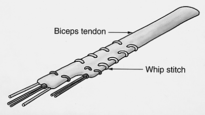
Fig. 1 Operative technique: one whip
stitch suture and two fibre wire sutures are inserted into the bicepital tendon
which has been trimmed to a chevron shape
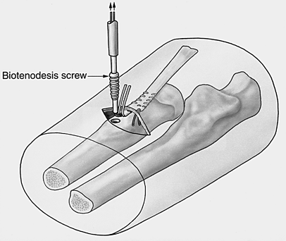
Fig. 2 Operative technique: the
Biotenodesis inserter containing the distal fibre wire suture is used to insert
a presized 5.5-mm screw into the radial tuberosity
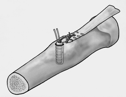
Fig. 3 Operative technique: the two
fibre wire sutures are tied together to appose tendon to bone
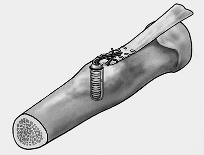
Fig. 4 Operative technique: the tied
fibre wire sutures are further tied to the whip stitch suture to allow further
apposition and support
On
the 1st day postoperatively the patient was placed in a hinged brace from
30–130° for 6 weeks. Return to unrestricted activity, including lifting,
was allowed over the next 6 weeks. There were no postoperative
complications and he regained full range of motion and strength by
3 months.
Discussion
Rupture
of the distal biceps tendon is a relatively uncommon injury and only around 300
cases have been reported in the literature [16].
The rupture typically occurs at the tendon insertion into the radial tuberosity
in an area of pre-existing tendon degeneration [16].
Surgical
intervention has been reported to achieve better results, especially with
restoring supination power. Current management tends to favour anatomical
reinsertion to the radial tuberosity in complete injuries, especially in active
and compliant young individuals desiring maximum return of elbow supination and
flexion power and endurance [1,
4,
12,
13,
14,
17,
20,
21,
22].
The
surgical management options include the single incision technique frequently associated
with suture anchor attachment [1,
9,
20,
24]
and the Boyd and Anderson double incision technique [6,
7].
The use of suture anchor attachment through a single limited anterior incision
has been shown not to be as stiff or strong as bone tunnel repairs in cyclic
loading and load-to-failure testing in vitro [3,
15].
The
minimal volar dissection needed for a limited anterior approach has been
associated with the reduced number of complications of synostosis and posterior
interosseous nerve injury [20,
22].
Bioabsorbable interference screws have been shown to be a safe and nonreactive
alternative to traditional metal interference screws for ACL graft fixation [11,
18,
19]
and have been shown to have a greater pullout strength when compared to suture
anchors. This absorbable screw is a valuable tool for the surgeon as it
minimises the problems of fixation loosening, migration, interference with
imaging studies and the potential requirement for later removal [2,
8].
In terms of primary fixation strength, the biodegradable interference screws
have been shown to be strong enough to allow accelerated rehabilitation and a
reasonable alternative to titanium interference screws [18,
19].
A
limited single anterior incision with Biotenodesis screw fixation is a safe and
effective alternate surgical option in the treatment of distal bicepital tendon
avulsions. The system allows for insertion via a limited exposure and provides
a strong fixation allowing early active motion and return to previous
activities as seen in our case.
References
|
1.
|
Aldridge JW, Bruno RJ, Straunch RJ,
Rosenwasser MP (2000) Management of acute and chronic biceps tendon rupture.
Hand Clin 16:497–503
 
|
|
|
|
|
2.
|
Barber FA, Deck MA (1995) The in vivo
histology of an absorbable suture anchor: a preliminary report. Arthroscopy
11:77–81
|
|
|
|
|
3.
|
Berlet GC, Johnson JA, Milne AD, Patterson
SD, King GJ (1998) Distal biceps brachii
tendon repair. An in vitro biochemical study of tendon reattachment. Am J
Sports Med 26:428–432
|
|
|
|
|
4.
|
Bernstein AD, Breslow MJ, Jazravi LM
(2001) Distal biceps tendon ruptures: a historical perspective and current
concepts. Am J Orthop 30:193–200
|
|
|
|
|
5.
|
Boileau P, Krishnan SG, Coste JS, Walch G
(2002) Arthroscopic biceps tenodesis: a new technique using bioabsorbable
interference screw fixation. Arthroscopy 18:1002–1012

|
|
|
|
|
6.
|
Boyd HB, Anderson LD (1961) A method for
reinsertion of the distal biceps brachii tendon. J Bone Joint Surg Am
43:1041–1043
|
|
|
|
|
7.
|
D Arco P, Sitler M, Kelly J,
Moyer R, Marchetto P, Kimura I, Ryan J (1998)
Clinical, functional, and radiographic assessments of the conventional and
modified Boyd-Anderson surgical procedures for repair of distal biceps tendon
ruptures. Am J Sports Med 26:254–261 Arco P, Sitler M, Kelly J,
Moyer R, Marchetto P, Kimura I, Ryan J (1998)
Clinical, functional, and radiographic assessments of the conventional and
modified Boyd-Anderson surgical procedures for repair of distal biceps tendon
ruptures. Am J Sports Med 26:254–261
|
|
|
|
|
8.
|
Lajtai G, Noszian I, Humer K, Unger F,
Aitzetmuller G, Orthner E (1999) Serial magnetic resonance imaging evaluation
of operative site after fixation of patellar tendon graft with bioabsorbable
interference screws in anterior cruciate ligament reconstruction. Arthroscopy
15:709–718
|
|
|
|
|
9.
|
Lintner S, Fischer T (1996) Repair of
distal biceps tendon using suture anchors and an anterior approach. Clin
Orthop 322:116–119
|
|
|
|
|
10.
|
McReynolds IS (1963) Avulsion of the
insertion of the biceps brachii tendon and its surgical treatment. J Bone
Joint Surg Am 45:1780–1781
|
|
|
|
|
11.
|
Morgan CD, Gehrmann RM, Jayo MJ, Johnson
CS (2002) Histological findings with a bioabsorbable anterior cruciate
ligament interference screw explant after 2.5 years in vivo. Arthroscopy
18:E47
|
|
|
|
|
12.
|
Morrey BF, Askew LJ, An KN, Dobyns JH
(1985) Rupture of the distal tendon of the biceps brachii. A biomechanical
study. J Bone Joint Surg Am 67:418–421
|
|
|
|
|
13.
|
Morrison KD, Hunt TR 3rd (2002) Comparing
and contrasting methods for tenodesis of the ruptured distal biceps tendon.
Hand Clin 18:169–178
|
|
|
|
|
14.
|
Norman WH (1985) Repair of avulsion of
insertion of biceps brachii tendon. Clin Orthop 193:189–194
|
|
|
|
|
15.
|
Pereira DS, Kvitne RS, Liang M,
Giacobetti FB, Ebramzadeh E (2002) Surgical repair of distal biceps tendon
ruptures: a biomechanical comparison of two techniques. Am J Sports Med
30:432–436
|
|
|
|
|
16.
|
Ramsey ML (1999) Distal biceps tendon
injuries: diagnosis and management. J Am Acad Orthop Surg 7:199–207
|
|
|
|
|
17.
|
Rantenen J, Orava S (1999) Rupture of the
distal biceps tendon. A report of nineteen patients treated with anatomic
reinsertion, and a meta-analysis of 147 cases found in literature. Am J
Sports Med 27:128–132
|
|
|
|
|
18.
|
Rupp S, Krauss PW, Fritsch EW (1997)
Fixation strength of a biodegradable interference screw and a press-fit
technique in anterior cruciate ligament reconstruction with a BPTB graft.
Arthroscopy 13:61–65
|
|
|
|
|
19.
|
Rupp S, Seil R, Schneider A, Kohn DM
(1999) Ligament graft initial fixation strength using biodegradable
interference screws. J Biomed Mater Res 48:70–74
|
|
|
|
|
20.
|
Sotereanos DG, Pierce TD, Varitimidis SE
(2000) A simplified method of repair for distal biceps tendon ruptures. J
Shoulder Elbow Surg 9:227–233
|
|
|
|
|
21.
|
Straunch RJ (2001) Techniques of distal
biceps tendon repair. Curr Opin Orthop 12:338–342
|
|
|
|
|
22.
|
Strauch RJ, Michelson H, Rosenwasser MP
(1997) Repair of rupture of the distal tendon of the biceps brachii. Review
of literature and report of three cases treated with a single anterior
incision and suture anchors. Am J Orthop 26:151–156
|
|
|
|
|
23.
|
Szymankiewickz J, Ramesh FT, Bunker T
(2003) Biotenodesis post: a novel technique for knotless arthroscopic cuff
repair. British Elbow and Shoulder Society, 14th Annual Scientific Meeting,
May 2003
|
|
|
|
|
24.
|
Verhaven E, Huylebroek J, Van
Nieuwenhuysen W, Van Overschelde J (1993) Surgical treatment of acute biceps
tendon ruptures with a suture anchor. Acta Orthop Belg 59:426–429
|
|
|
|