
Arthroscopic Insertion of a Biologic Rotator Cuff Tissue Augmentation After Rotator Cuff Repair
Authors: Seldes RM, Abramchayev I
References: Arthroscopy. January 2006 (Vol. 22, Issue 1, Pages 113-116)
Abstract
Tissue augmentation has been used to reinforce repairs of massive chronic rotator cuff tears. This report describes a new technique for arthroscopic insertion of a biologic rotator cuff tissue augmentation. Double-loaded suture anchors are needed for the technique. One suture is used for the rotator cuff repair and 1 suture is used for the tissue augmentation. The sutures for the tissue augmentation are left exiting through accessory portals. Two free sutures are then passed through the repaired rotator cuff, 1 anteromedially and 1 posteromedially via the anterior and posterior cannulae, respectively. One limb from each of these 4 sutures is retrieved through the lateral portal. Each of the 4 limbs exiting through the lateral portal are then passed through the corresponding corner of the tissue augmentation and Mulberry knots are tied in each suture limb, on top of the tissue augmentation. The suture limbs of the 4 sutures exiting through the anterior and posterior cannulae and accessory portals are pulled simultaneously to reduce the tissue augmentation down onto the repaired rotator cuff. A trocar is used to help push the tissue augmentation through the lateral cannula. The Mulberry knots from each of the sutures are sequentially retrieved and tied arthroscopically to secure the tissue augmentation.
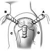
Figure 1. Anteromedial and posteromedial sutures exiting through anterior and posterior cannulae. Anterolateral and posterolateral sutures from suture anchors exiting through accessory portals.
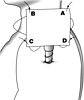
Figure 2. One limb from each of the 4 suture pairs is passed through the respective corner of the tissue augmentation and Mulberry knots are tied.
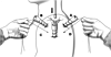
Figure 3. Tissue augmentation in reduced into shoulder through the lateral cannula.
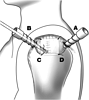
Figure 4. Mulberry knots are retrieved and arthroscopic knots are tied on all 4 corners of the tissue augmentation.
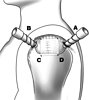
Figure 5. Final repair with tissue augmentation.
