
MRI scan
Print/Download Patient Information Leaflet (pdf)
MRI Scans (short for Magnetic Resonance Imaging) is a powerful imaging technique which exploits the magnetic properties of hydrogen atoms within the body. This imaging technique uses magnetic fields and radio waves and does not employ ionizing radiation which normal x-rays use.
MR scanning does not use x-rays and there are no known harmful effects. MR scans show soft tissues such as muscles, ligaments, tendons and cartilage extremely well. CT scans and x-rays are better for viewing bones. Since most sports injuries involve the muscles, tendons and ligaments MR scanning is extremely useful.
When looking at soft tissue structures within a joint MR arthrograms are useful for detection of ligament tears and cartilage abnormalities. This involves a small injection of special dye into the joint before the MR scan. The dye makes it much easier to see the joint structures. See video below.
We also use special views of the shoulder to better demonstrate sporting injuries such as small rotator cuff tears, Labral tears, SLAP tears and Throwers shoulder. These are called ABER views. Because this is so specialized and unique, we use the skills of Radiologists specially trained in this method to supervise and interpret these images.
 |
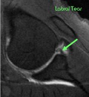 |
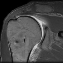
| Usual MRI with arm by side | ABER view with arm above head - demonstrating a tear not seen on usual MRI |
MRI Arthrogram Injection:
